Calcium Imaging at Karolinska Institutet, Sweden
Dr. Wiktor Phillips
Department of Women's and Children's Health, Karolinska Institutet, Sweden
Background
Dr. Wiktor Phillips is primarily interested in observing the pattern generation of respiratory rhythms in mammals through the study of the pre-Bötzinger complex in the medulla of the brainstem, and whether these patterns are affected by birth and related to respiratory dysfunction in newborns.
This group investigates pattern generation in these complexes through in vitro study of mouse brain slices, either taken acutely or organotypically cultured for several weeks. These slices are subject to both single-cell patch clamping and whole network calcium imaging, often over long experiments where drugs can be administered, onset of activity can be monitored, and sub-populations of neurons and astrocytes can be functionally compared.
A variety of fluorophores are used for calcium imaging, including Cal-590™ AM for red and Fluo 8® AM/Cal-520® AM for green, in combination with patch clamping.

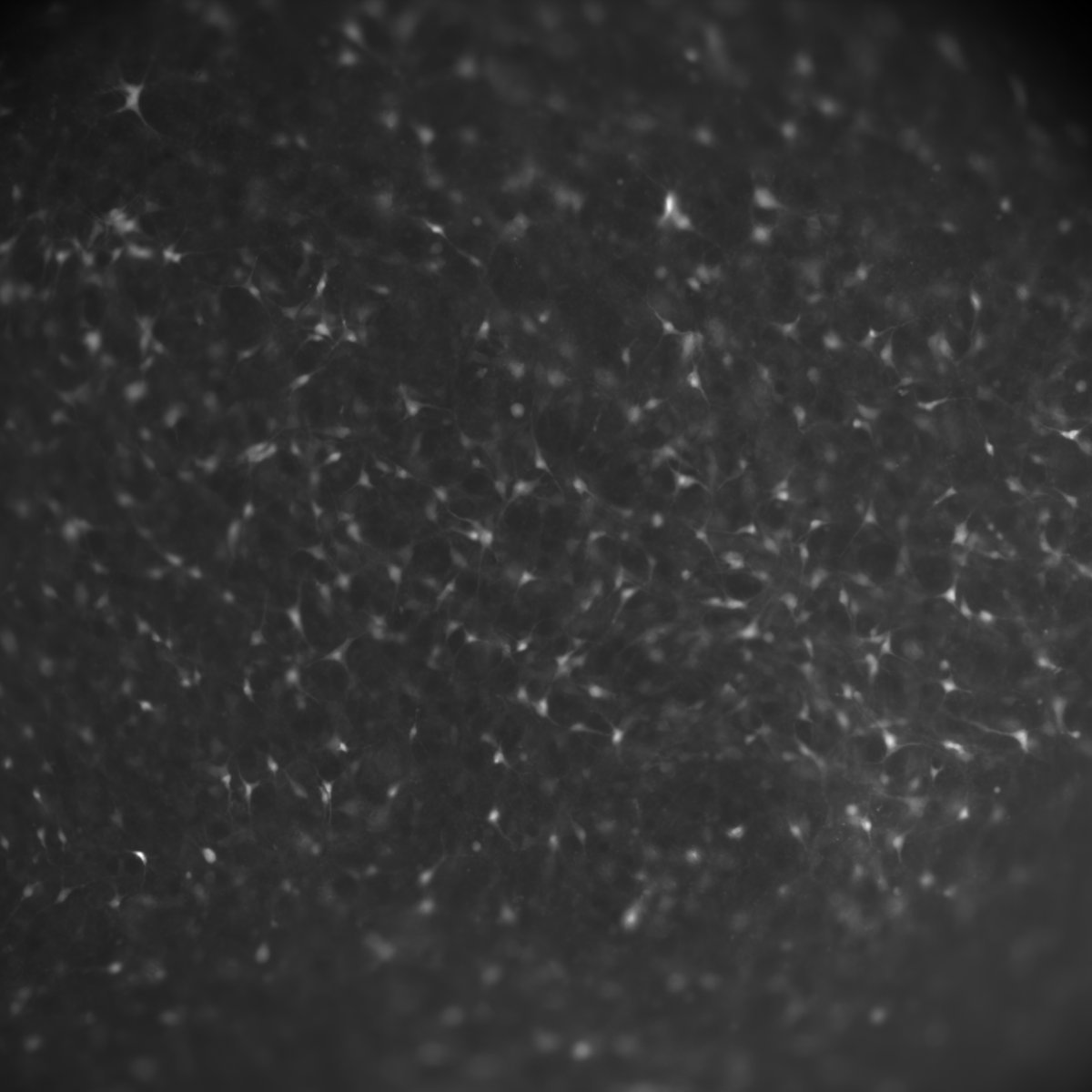
Figure 1: An image taken with the Prime 95B, showing neural cells labeled with Cal-590AM
(a red calcium dye) in an acute slice of brainstem tissue.
Challenge
When imaging sub-cellular functional dynamics via patch clamping as well as whole-network calcium imaging with numerous functional populations of neurons and astrocytes, it is necessary to have highly sensitive imaging equipment with high framerates and a wide field of view. In addition, when calcium imaging brain slices there can be issues with light scattering depending on the age of the slice, and time lag while the calcium fluorophore dyes bind to the sites of interest.
As Dr. Phillips uses a combination of different electrophysiological and imaging hardware it became necessary to use more customizable operating systems than Windows, namely Linux. Some imaging and electrophysiological software are locked to certain operating systems and not customizable, but in Linux it is possible to write custom scripts to support specific applications, even synchronizing between entirely different pieces of hardware. By using Linux experiments become far more flexible and optimizable, but as many products are only supported in Windows, the switch to Linux can be inconvenient.
We barely need any light and we still get a great image [with the Prime 95B].
Dr. Wiktor Phillips
Solution
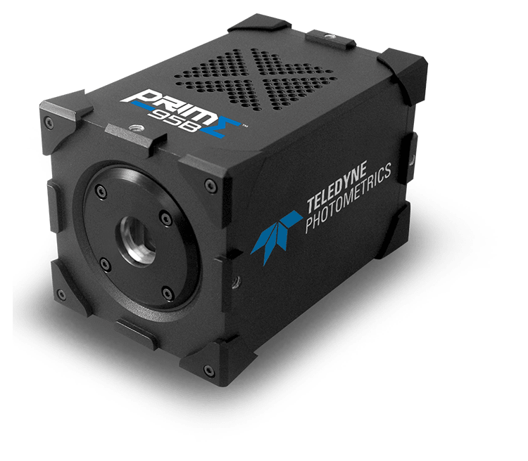
The Prime 95B offers both a hardware and software solution, being fully supported in Linux and featuring the optimal hardware specifications for imaging both sub-cellular and whole-network brain slices.
The large sensor of the 95B allows for high-speed images to be acquired using a region of interest (ROI) without sacrificing the required field of view when working with single cell patch clamping, as well as the use of the full-frame sensor for whole network calcium imaging. The Prime 95B offers a flexible solution for any scale or length of experiment and excels in low light conditions. As Dr. Philips told us "We barely need any light and we still get a great image".
The 95B is well supported in Linux which allows for flexible software customization, with the PVCAM Linux driver also tested frequently with Ubuntu: "some software is locked to Windows but with Photometrics, I could write custom scripts and interfaces for the Prime 95B camera in Linux and pair recordings from whole-network calcium imaging and patch clamping simultaneously".

Learn More About The Prime 95B