Intravital NIR Imaging
Dr. Epameinondas Gousopoulos (MD/Ph.D.), Dr. Stefan Wolf
Division of Plastic and Hand Surgery, University Hospital Zürich, Switzerland
Background
The group of Prof. Gousopoulos at the University Hospital Zurich is focused on researching lymphedema, a condition where lymphatic system dysfunction results in swelling in parts of the body. The group has established a mouse model in order to investigate this disease.
Postdoc Dr. Stefan Wolf told us more about his research, "We use surgery to remove lymphatic vessels in the mouse tail, this introduces lymphedema and causes swelling in the mouse tail. We then image the lymphatic vessels within the tail using an intravital microscope setup."
"We inject a lymphatic-specific near-infrared (NIR) fluorescent tracer dye into the tail tip and image the flow down the tail, visualizing the capillaries and lymphatic collectors. This helps us understand the underlying mechanisms, as well as screen for new pharmacological compounds which may influence the onset of lymphedema or protect the lymphatic vessels."

Figure 1: Flow of a NIR dye through a section of a mouse tail, imaged with the Prime BSI Express.
Hairs, capillaries, and lymphatic vessels are all visible within the tail.
Challenge
Intravital imaging involves imaging large live organisms, in this case, mice. Breathing and other small movements can decrease image quality, and small magnifications are needed to capture large areas of the sample. In this case, imaging uses magnifications between 4x and 12x, requiring a camera with a small pixel in order to best match Nyquist sampling and get good resolution.
Low magnifications also allow for a large field of view, so a camera with a large sensor is also suitable, which can also image at a video rate in order to capture dynamic movements of the dye through the lymphatic system within the tail.
In addition, as the lymph networks are beneath the skin, NIR wavelengths are used to penetrate below the tail surface and image the dye within. This requires a camera with high sensitivity and quantum efficiency in NIR wavelengths (>700 nm), in order to best capture the signal.
I am very pleased with the [Prime BSI Express], I’ve never seen a camera that can image such a tiny signal with hardly any noise, it’s everything we wished for from a camera and perfect for us!
Dr. Epameinondas Gousopoulos (MD/Ph.D.), Dr. Stefan Wolf
Solution
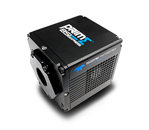
The Prime BSI Express is a compact yet powerful sCMOS camera with a small pixel, large sensor and sensitivity far into the NIR range, making it an ideal solution for this application.
Dr. Wolf told us more about his experience with the Prime BSI Express, "We combined the camera with a Zeiss microscope for intravital imaging, it worked really well with Zen imaging software, and the camera instantly connected and we had no problems at all."
"We definitely needed the sensitivity of the sensor in order to capture even faint traces of the dye and have a much better range of data. I've never seen a camera before that can image such a tiny signal with hardly any noise, it's everything we wished for!"
"We might also need the speed of the Prime BSI Express for other projects, such as imaging blood flow within capillaries at wound sites. We wanted the perfect solution for our experiments now and for the future."

Learn More About The Prime BSI Express
Download This Customer Story