Novel Hyperspectral Imaging
Prof. Silas Leavesley
Department of Chemical and Biomolecular Engineering, University of South Alabama, US
Background
The interdisciplinary laboratory led by Prof. Silas Leavesley and Dr. Thomas Rich is working to develop novel hyperspectral imaging systems for microscopy and endoscopy. Using rapidly controllable light sources with precise spectral selection they aspire to be able to increase the number of individual sensors and probes detected concurrently. This will allow measurements of spatial and temporal fluctuations in various signal transduction cascades in cells, tissues, and organisms.

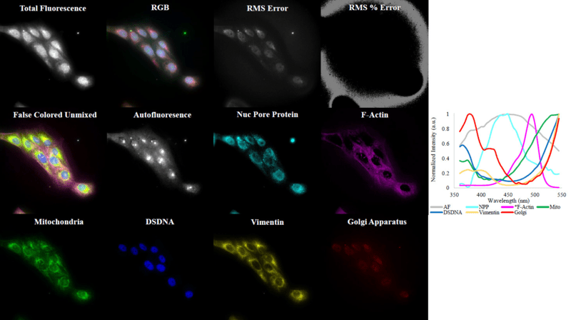
Figure 1: Spectral excitation-scanning images of a 6-label slide (Abberior with autofluorescence). Excitation wavelengths were sampled over a range of 360-540 nm, at a 5 nm increment, using a Xe arc lamp and Sutter VF 5 filter assembly equipped with Semrock VersaChrome filters, while image data were acquired using a 555 nm long-pass emission filter and Prime 95B sCMOS camera. The raw fluorescence signal can be visualized as Total Fluorescence (the summed intensity across all wavelength bands) or RGB false-colored (a visualization of 3 selected and false-colored wavelength bands). A spectral library was constructed from single-label controls (plot at right). The spectral image cube was then unmixed using non-negatively constrained linear unmixing. The residual (unaccounted for) signal is displayed as both total signal, called root-mean-square (RMS) Error, and the relative error, as a percent of the RMS signal in each pixel, called RMS % Error. Linearly unmixed images were false colored for visualization and were also merged, shown by the False Colored Unmixed.
Challenge
Acquisition of hyperspectral image data on timescales relevant to biochemical and cellular processes requires rapid selection of precise excitation wavelengths and rapid, low-noise image acquisition of resulting excitation-scanning spectral image stacks.
Given the aim of acquiring 1-5 spectral image data sets of 30 wavelength bands each second, a fast, sensitive low-noise camera is an absolute requirement to generate data of sufficient signal-to-noise ratio for interrogating cellular processes.
Previous work using widefield imaging with EMCCD cameras was successful, but the field of view and slow readout speed of EMCCD detectors limited the spatial and temporal sampling of the desired measurements. Now, Prof. Leavesley's lab is aiming to acquire similar spectral data using spinning disk confocal microscopy to allow 3D, timelapse, and spectral imaging capabilities.
The large pixel size, larger sensor format, and high-speed readout of the Prime 95B allows for high speed spectral imaging when performing sequential wavelength band scanning.
Silas Leavesley
Solution
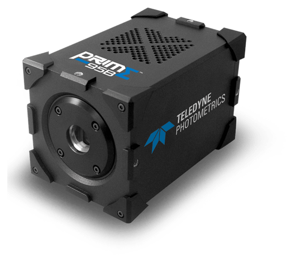
The Prime 95B is a highly sensitive CMOS camera with a large field of view, matching a near-perfect 95% quantum efficiency with low read noise for a high signal-to-noise ratio when imaging.
Prof. Leavesley told us about his experience with the Prime 95B CMOS, "The Prime 95B, with its high quantum efficiency, large FOV, high readout speed, and low readout noise allowed us to gather spectral cubes at N spectral volumes per second, image cubes at >1 Hz frequencies, and will be a key component in developing future 5-dimensional imaging approaches."

Learn More About The Prime 95B