Ultra-High-Sensitivity emICCD Cameras Enable Diamond Quantum Dynamics Research
Introduction
Interest in the various crystal defects found within diamonds is growing quickly amongst physicists and biologists worldwide. This increasing attention is attributable in no small part to the ability of such defects, when embedded in nanocrystals, to function as single-photon sources or as highly photostable, low-cytotoxicity fluorescent biomarkers.1
One of these defects, the nitrogen-vacancy (NV) center, can be utilized to detect and measure local magnetic2 and electric fields,3 a capability based on the quantum mechanical interactions of the defect's spin state. The ongoing study of NV centers holds fantastic promise for a broad range of advanced applications, including ion concentration measurements,4 membrane potential measurements,5 nanoscale thermometry,6 and single-spin nuclear magnetic resonance.7
Recently, researchers at the Université de Sherbrooke in Québec, Canada, led by Ph.D. student David Roy-Guay and supervised by professors Denis Morris and Michel Pioro- Ladrière, employed a scientific emICCD camera from Princeton Instruments to image the quantum state of NV centers in diamond over a wide surface (100 μm x 100 μm) on a pixel basis. Their work on diamond quantum dynamics is the focus of this application note.
Nitrogen-Vacancy Basics
Nitrogen-vacancy centers in diamond show exceptional light-to-spin properties that can be exploited in quantum information,8 magnetic resonance imaging, and entangled photon sources.9 Embedded in the rigid structure of diamond, the radiative defect is composed of a single substitutional nitrogen atom adjacent to a carbon vacancy (see Figure 1a) freeing two electrons with a spin, a quantum property. Combination of their spin forms a triplet state,10 sensitive to the external magnetic field applied. Usually, even the smallest change in the magnetic field induced by other spins in a material affects the spin properties of a defect. In the case of the NV centers, such modulations are limited by the low spin-phonon coupling and the low concentration (~1%) of other spin species, resulting in the preservation of the spin triplet coherence even at room temperature.11 Combined with the ability to read out the spin state optically, NV centers make outstanding magnetic field sensors with nanoscale sensitivity.12
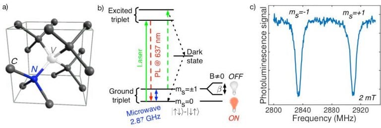
Figure 1: (a) Lattice structure of diamond, including the NV center.
(b) Energy-level structure of the NV center. (c) Optically detected
magnetic resonance (ODMR) of the NV center for a 2 mT external
magnetic field. Courtesy of David Roy-Guay, Université de Sherbrooke.
A closer look (see Figure 1b) at the energy levels of the NV center shows the close relationship between light and spin property. The ground triplet state of the NV center is linked to the excited state by a radiative transition at 637 nm. Its spin projection is either 0 (symmetric state, |0>) or ±1 (both spins up or down, |±1>), split by 2.87 GHz. Following excitation with laser pulses at 532 nm, the NV center initially in state 0 will re-emit red photons at 637 nm. On the other hand, if the initial state is ±1, it will de-excite via a non-radiative transition to the 0 state on a timescale of approximately 300 nsec. Consequently, the intensity of the red light collected depends on the state, and the NV center can be initialized by sending a short (µsec) laser pulse. Upon application of an external magnetic field along the NV quantization axis, the ±1 state is split by quantity β (28 MHz/mT), proportional to the magnetic field.
By sending microwaves in an optically detected magnetic resonance (ODMR) experiment, the spin state can be manipulated (see Figure 1c). Recording the photoluminescence while scanning the frequency of the microwaves, the amplitude and orientation of an unknown magnetic field can be extracted via measurement of the distance between the two spin states, which appear as dips in Figure 1c. Consequently, one sees that narrow resonance lines improve the magnetic field sensitivity.
Coherent Control of the NV Center State
The experimental setup used by David Roy-Guay to measure the ODMR is shown in Figure 2. The green laser goes through a double-pass acousto-optical modulator (AOM) to generate laser pulses for initialization and readout of the NV centers. A 60X plano-convex microscope lens focuses the laser on the surface of the NV-containing CVD diamond and the emitted light is collected by a PI-MAX4:512EM emICCD camera.
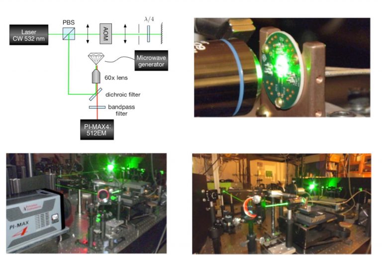
Figure 2: Experimental setup for NV center imaging. Laser pulses
are generated by the AOM in a double-pass configuration and
sent to the sample with a polarizing beam splitter (PBS).
Courtesy of David Roy-Guay, Université de Sherbrooke.
In the spin-manipulation experiment shown in Figure 3a, an external field (10 mT) is applied to split the levels ±1 into the four possible orientations of the NV in the lattice structure. The resonant microwave pulses for the orientation at 2751 MHz are amplified and sent through a photolithographically defined wire on the surface of the diamond - these pulses' widths are stepped in-between the acquisitions of the camera.
Synchronization of the microwave generator, the AOM, and the emICCD camera is achieved by operating the camera in its external trigger mode and counting the number of frames out-put by the 'logic out' output.
Because of the fast repolarization of the spin state to 0, the gate must be precisely aligned with the laser readout pulse. This critical alignment is easy to perform via the sequential gating function of the PI-MAX4:512EM camera, which moves the gate pulse relative to the trigger pulse. Once the gate pulse is set at the beginning of the readout laser pulse, microwaves are applied with a variable time τ for coherent control of the NV spin state (see inset Figure 3a). The quantum dynamics of an NV ensemble caught on a single pixel (blue curve Figure 3a) or averaged over a 10×10 pixel region (red curve) show excellent signal-to-noise ratio.
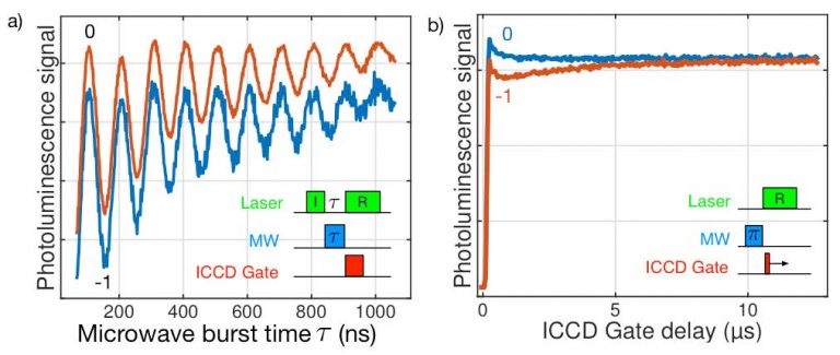
Figure 3: (a) Rabi oscillations of an NV ensemble on an individual pixel
(blue) and 10×10 pixel region (red). Inset: pulse sequence for the laser
initialization (I) and readout (R) of the NV centers. Microwave (MW) pulses
are applied between the laser pulses.
(b) Photoluminescence readout of the NV center state. Maximum contrast
is obtained by integrating the signal over the first µsec of the laser
pulse. Courtesy of David Roy-Guay, Université de Sherbrooke.
The oscillations show that the spin state can be flipped from 0 to -1 by applying a 150 nsec pulse, corresponding to a π-pulse. Curve decay is due to relaxation: over 1 µsec, the ensemble of NV centers probed interacts with other spins in the diamond lattice so that their quantum state can no longer be manipulated coherently with high fidelity. Once the π pulse duration has been determined, the spin repolarization dynamics can be measured by preparing the NV in the 0 or -1 state and scanning the gate across the readout pulse (see Figure 3b).
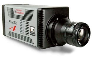
Figure 4: The PI-MAX4:512EM from Princeton Instruments
gives users the benefits of both an intensified CCD (ICCD)
camera and an electron-multiplying CCD (EMCCD) camera.
This fiber-optically bonded Princeton Instruments PI-MAX®4 camera system provides <500 psec gate widths using standard fast-gate intensifiers while preserving quantum efficiency. Its integrated SuperSynchro timing generator allows camera users to set gate pulse widths and delays under GUI software control, and significantly reduces the inherent insertion delay (~27 nsec).
Complete control over all PI-MAX4:512EM hardware features is simple with the latest version of Princeton Instruments' LightField® data acquisition software (available as an option). Precision intensifier gating control and gate delays, as well as a host of novel functions for easy capture and export of image data, are provided via the exceptionally intuitive LightField user interface. The PI-MAX4:512EM uses a high-bandwidth (125 MB/sec or 1000 Mbps) GigE data interface to afford camera users real-time image transmission. This interface supports remote operation from more than 50 meters away.
Future Directions
The use of a scientific emICCD camera from Princeton Instruments has allowed researchers at the Université de Sherbrooke to image the quantum state of nitrogen-vacancy centers in diamond over a wide surface (100 µm x 100 µm) on a pixel basis. Observing and studying the effects of various magnetic systems on the optically detected magnetic resonance, ranging from functionalized biological samples to magnetic domains in materials, will be possible by tracking the position of the ODMR peaks down to 1 Gauss variations.
Furthermore, higher-sensitivity magnetometry can be achieved by applying spin-echo pulse techniques enabled by the emICCD camera's precision gating features. Such a capability is key to the development of a new scientific tool based on diamond technology, one which will provide unique insights for biology, medicine, chemistry, physics, and geology.
Acknowledgments
Princeton Instruments thanks David Roy-Guay (Université de Sherbrooke) for his invaluable contributions to this application note. The research team at the university would like to acknowledge funding from FRQNT and CIFAR.
Resources
To learn about the latest diamond quantum dynamics research being conducted at the Université de Sherbrooke, please visit:
http://www.physique.usherbrooke.ca/~piom2101/index.php?sec=accueil&lan=EN
References
- Schirhagl R. et al. Nitrogen-vacancy centers in diamond: nanoscale sensors for physics and biology. Annu. Rev. Phys. Chem. 65, 83-105 (2014).
- Maze J.R. et al. Nanoscale magnetic sensing with an individual electronic spin in diamond. Nature 455, 644-647 (2008).
- Dolde F. et al. Electric-field sensing using single diamond spins. Nat. Phys. 7, 459-463 (2011).
- Hall L.T. et al. Monitoring ion-channel function in real time through quantum decoherence. Proc. Natl. Acad. Sci. U.S.A. 107, 18777-18782 (2010).
- Hall L.T. et al. High spatial and temporal resolution wide-field imaging of neuron activity using quantum NV-diamond. Sci. Rep. 2, 401 (2012).
- Toyli D.M. et al. Fluorescence thermometry enhanced by the quantum coherence of single spins in diamond. Proc. Natl. Acad. Sci. U.S.A. 110, 8417-8421 (2013).
- Mamin H.J. et al. Nanoscale nuclear magnetic resonance with a nitrogen-vacancy spin sensor. Science (80-) 339, 557-560 (2013).
- Bernien H. et al. Heralded entanglement between solid-state qubits separated by three metres. Nature 497, 86-90 (2013).
- Togan E. et al. Quantum entanglement between an optical photon and a solid-state spin qubit. Nature 466, 730-734 (2010).
- Gali A. et al. Theory of spin-conserving excitation of the N-V-center in diamond. Phys. Rev. Lett. 103, 1-4 (2009).
- Balasubramanian G. et al. Ultralong spin coherence time in isotopically engineered diamond. Nat. Mater. 8, 383-387 (2009).
- Balasubramanian G. et al. Nanoscale imaging magnetometry with diamond spins under ambient conditions. Nature 455, 648-651 (2008).
Further Reading
Ultra-Low-Light Imaging
in Quantum Research
Find out about the different types of camera
technologies that can be used for quantum
research,their advantages and their limitations.
emICCD Cameras for Using
Trapped Ions in Quantum Research
Find out how researchers from Germany created a
nano-heat engine with only a single ion,
and observed it using an emICCD.
<500 Picosecond Gating for
Atmospheric Pressure Plasma Jets
An application note providing an overview of the
experimental setups for atmospheric pressure
plasma jets alongside relevant imaging technology.
