Neural Vascular Imaging
Prof. Dave Attwell
University College London
Background
The Attwell lab is interested in understanding the interactions that occur between neurons, glial cells and the vasculature of the brain through the use of electrophysiology and imaging techniques.
For years it was believed that brain blood flow, which provides the energy used for neural computation, was controlled solely by constriction and dilation of arterioles in the brain. However, the Attwell lab have since shown that contractile cells called pericytes, located at 30-micron intervals along brain capillaries, also play a major role (Hall et al., 2014, Nature 508, 55; Mishra et al. 2016, Nature Neuroscience 19, 1619).
Capillaries are extremely small (around 4-5 microns in diameter) and therefore red and white blood cells need to change shape in order to pass through them. It is these changes in shape that the Attwell group are interested in imaging

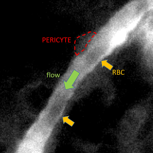
Figure 1 Pictures taken 1 ms apart with the Prime BSI, showing the passage along a
capillary (in an anaesthetised mouse brain) of red blood cells (RBCs; hazy black
objects in the middle of the capillary, with orange arrows pointing at them - the
bottom RBC moves out of the field of view during the time between the pictures). The
Attwell group are interested in how the RBCs interact with, and have their movement
affected by, the processes of contractile cells called pericytes which sit on the capillary
wall (a pericyte is outlined in red).
Challenge
The problem with imaging such changes of red and white blood cell shape is that the cells themselves move very quickly (~1mm/sec). They are also only 5 microns across, so they pass any given point in only a few milliseconds. They also change shape very quickly. Consequently, to capture images of the changes of shape, a rapid and high sensitivity imaging camera is needed.
The camera has allowed us to acquire images that would otherwise be impossible to obtain, providing a basis for future analysis of how the properties of capillaries, and of red and white blood cells, determine brain blood flow.
Dave Attwell
Solution
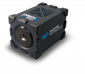
The Attwell lab required a high sensitivity and rapid acquisition camera, and thus are now using the Teledyne Photometrics Prime BSI with their imaging system.
Prof. Attwell told us that, "Based on prior interactions, we selected Teledyne Photometrics as a reliable source of information on this and ended up purchasing a Prime BSI for our imaging demands."
Prof Attwell went on to say that, "The Prime BSI is ideal for our purposes. By aligning a capillary with the camera image x-axis and choosing a smaller field of view in the y-axis, we can acquire images at ~1kHz, allowing us to image changes of the shape of the cells as they pass any given point in the capillary network. The camera has allowed us to acquire images that would otherwise be impossible to obtain, providing a basis for future analysis of how the properties of capillaries, and of red and white blood cells, determine brain blood flow".

Learn More About The Prime BSI