Calcium Imaging at Medical University of Vienna
Prof. Michael J.M. Fischer, PhD
Institute for Molecular Physiology, Medical University of Vienna
Background
Dr. Fischer's research addresses nociception, the nervous system's response to harmful or potentially harmful stimuli, with focus on the peripheral nervous system. A particular interest is the role of transient receptor potential (TRP) channels, which play an important role in sensing pain.
Typically, imaging experiments in the Fischer lab involve the use of fluorescent indicators in cultured primary neurons or cell lines. Sensors for calcium ions (Ca2+) and voltage are used as activity readouts for spontaneous and induced signal transduction - with sensors being either ratiometric or non-ratiometric.

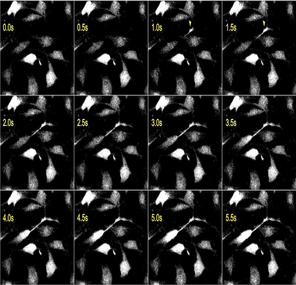
with the Photometrics Prime BSI. Upon application of a receptor agonist,calcium influx started in a cellular extension (yellow arrowheads),
followed by a spreading wave that reachesall cytosolic areas.Cells were stained with Fluo-8 and excited at 455 nm using an Omicron
LED HUB.Thereceptor distribution is compared to the start of the initiation of the calcium wave by subsequent confocalimaging of immunocy to chemistry.
Challenge
A typical experiment performed by the group uses a chemical cue to generate rolling Ca2+ waves along a cellular extension which spread across the cell. Depending on the traveling speed, this requires a very high sampling rate. High-speed image acquisition, as well as structural-functional correlation, present important imaging challenges faced by the lab.
For conventional imaging techniques, observation of these Ca2+ waves requires a compromise between the acquisition speed necessary to obtain any meaningful kinetics and the low magnification needed to achieve substantial output. This is especially important when obtaining precise Ca2+-concentrations using ratiometric sensors such as Fura-2, which require switching between two excitation wavelengths.
In the best-case scenario, the Ca2+ signals will need to be imaged at close to 1000 frames per second (fps) and triggering through a software solution can be too slow to achieve this.
The direct control of other devices by TTL output allowed reliable high-speed measurements triggered by the camera without a separate real-time controller. The combination of high sensitivity and the ability for fast measurements making use of this sensitivity became very useful.
Michael , J.M. Fischer, PhD
Solution
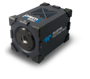
The Prime BSI enables the Fischer lab to image the progression of Ca2+ waves across neurites with unprecedented speed, accuracy, and precision.
Even ratiometric Fura-2 imaging can be achieved with an effective speed of up to 460 fps (>920fps, alternating 340/380 nm excitation) using the SMART (Sequenced Multiple Acquisition in Real Time) streaming feature implemented on Photometrics Prime cameras.
SMART streaming allows the camera to directly hardware-trigger up to 4 individual excitation channels with individually adjustable exposure times and intensities.
When imaging at these speeds, every single photon counts. The Prime BSI, with 95% quantum efficiency and very low read noise, produces results with an excellent signal-to-noise ratio and a very high dynamic range, which makes the camera the perfect match for the Fischer lab's application.

Learn More About The Prime BSI