Plant Stress Calcium Imaging
Prof. Rob Roelfsema
MOLECULAR PLANT-PHYSIOLOGY AND BIOPHYSICS, UNIVERSITY OF WURZBURG, GERMANY
Background
Prof. Rob Roelfsema is studying plant signaling and transport, typically using fluorescent ion sensors to see changes in calcium concentrations during certain conditions. Prof. Roelfsema told us more, “We use Arabidopsis, tobacco, and the Venus flytrap as models to show people that plants are actually sensing organisms, and they are more alive than most people realize.”
“We image the leaves of plants that express an ion sensor, often a calcium sensor, and then we apply stress to the leaf and observe the calcium signaling, which typically moves like a wave through the leaf.”
“With the Venus flytrap we stimulate the trigger hairs twice within 10 seconds and this activates the trap, and with the Arabidopsis, it’s more brutal, we are squeezing or burning the leaves to look at the strong stress signals. It looks brutal but if you think of a caterpillar eating the leaf, it is also brutal, and we want to see what signals the plant sends to defend itself.”
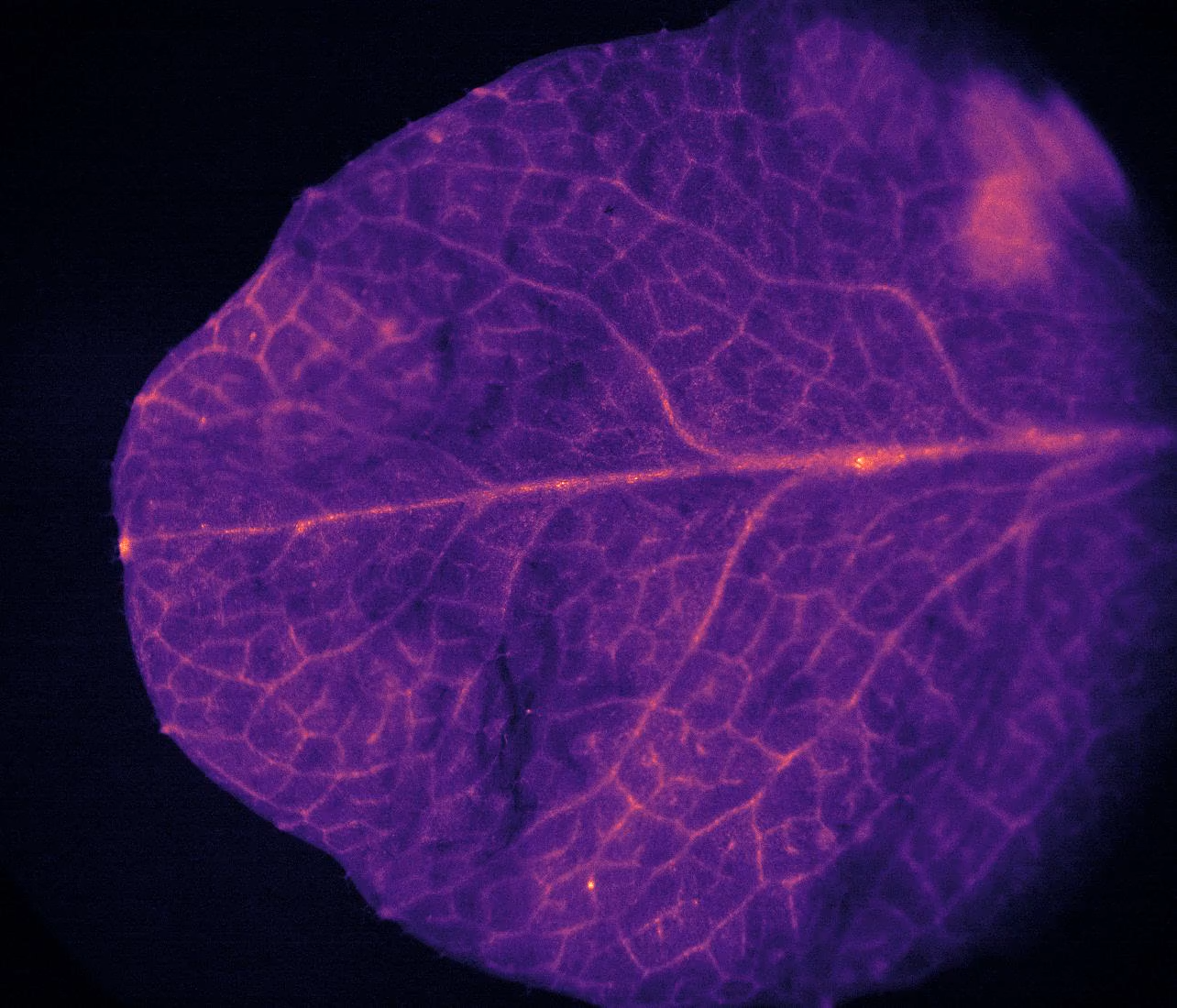
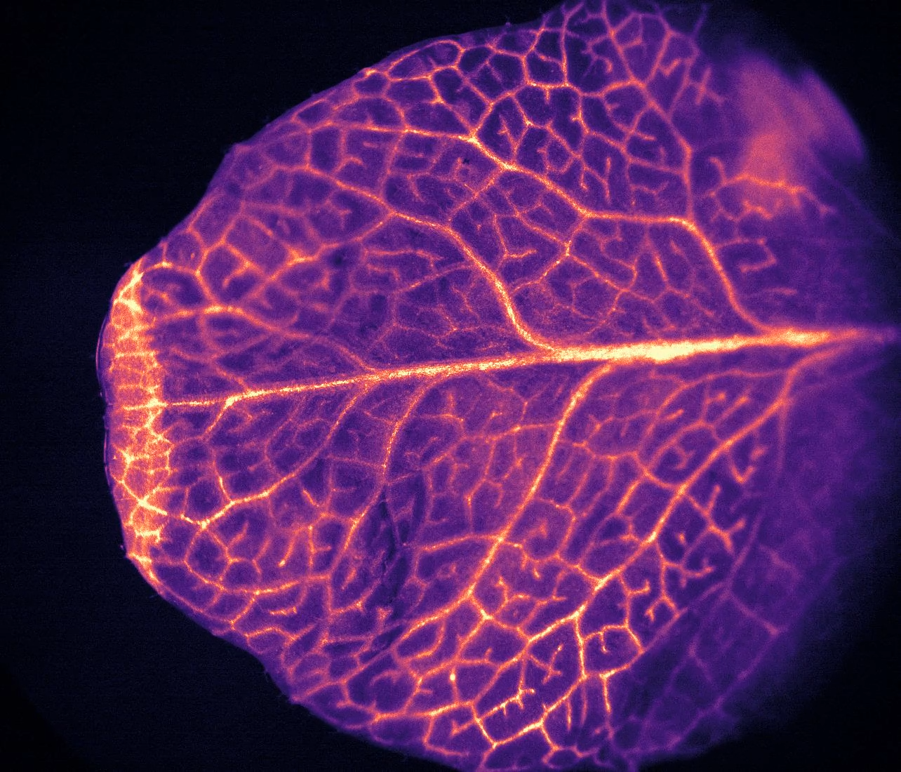
5x-sped-up_RGECO_04_Arabidopsis_on-fire_250ms_100ms_2x2binning_MMStack_Pos0.ome_.mp4


Figure 1:Calcium imaging of plant models using the Retiga E7 CMOS camera. These images show an Arabidopsis leaf before and after being burned, showing calcium signals propagating in waves through the leaf due to stress from heat and damage. A video of this calcium activity can be seen in the link above the images.
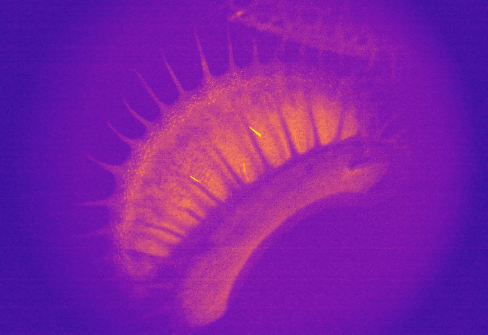
Fly-Trap-GCaMP6_1_MMStack_Pos0.ome-1.mp4

Figure 1: Calcium imaging of plant models using the Retiga E7 CMOS camera. Image shows a Venus flytrap after being stimulated at one of the trigger hairs (seen as bright yellow structures in the trap), and just about to close. Full video of stimulation and closing can be see in the link above the image.
Challenge
Calcium signals are rapid and dynamic, this requires an imaging system that can operate at high speeds while still having the sensitivity to capture the signal.
Prof. Roelfsema described his imaging challenges, “We are imaging calcium waves so we need to capture at about 100 milliseconds or less, but the signal is also in the lower level, and if we reduce the exposure time too much, we drop below the detection level. We also need to consider that plants are very sensitive to light intensity, we are using a UV lamp, and if we are focusing on a single cell the light intensity can be very high.”
Another factor is the field of view, these calcium waves can spread across a whole leaf or section of plant tissue, and the greater the area that can be imaged, the more relevant information can be obtained.
With the Retiga E7 we can image these calcium signals moving in waves through larger plant tissues, that’s something we couldn’t do before.
Prof. Rob Roelfsema
Solution
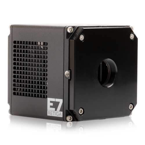
The Retiga E7 CMOS camera is an ideal entry-level solution for calcium imaging, with the ability to image at 50 fps with low read noise, all with a large field of view.
Prof. Roelfsema explained his experience with the Retiga E7 camera, “We can image the signal moving through the whole leaf tissue like a wave and we couldn’t do that before, our older cameras were much too slow. That’s why we bought this camera, to be able to image these calcium signals in larger tissues at once.”
“There have been cameras that were capable of getting the signal, but they were not affordable, now the Retiga E7 is definitely more in the range of what I could easily afford. Also, setup of the camera in MicroManager was easy.”
