Live Cell Imaging
Jessica Kehrer, Dipl. Ing. & Prof. Friedrich Frischknecht
Frischknecht lab, Centre for Infectious Diseases, Parasitology
Heidelberg University Medical School
Background
The Frischknecht lab aims to understand transmission of the malaria parasite between host and mosquito. The lab focuses on the two motile stages of the life cycle - the ookinete and the sporozoite using the rodent model parasite Plasmodium berghei. The movement of this parasite is particularly interesting because the cells are able to move at very high speed without changing their shape.
For their standard experiments, they are using reverse genetics to either remove proteins or to label them with fluorescent markers followed by characterization of the resulting parasite line with state of the art microscopy techniques.

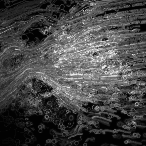
Figure 1. Maximum intensity projection of Plasmodia released from salivary glands.
Cells were labelled with GFP. Imaging frequency was 250 fps.
Challenge
Motility can be studied using sporozoites freshly isolated from the salivary gland of infected mosquitoes. Wild-type sporozoites generally show a circular movement which proceeds at a speed of 1.5-2 µm/s for up to one hour.
The current imaging solution in the Frischknecht lab is a front illuminated CCD sensor with a very limited field of view (FOV) and low sensitivity. Sensitivity limitations result in long exposure times which in turn make the analysis of the obtained data - involving tracking of individuals and studying their motion - difficult. Typically, exposure times of 300-400 ms or more for the individual channels is common. Therefore, it is currently impossible to image the localization of weak fluorescent proteins.
The relatively low FOV also results in a lower sample throughput, reducing the quality of the statistical information - especially for mutants with low infection rates.
If sensitivity could be increased, the temporal resolution could also be improved. This would be crucial for improving tracking of the individual parasites, producing higher quality data that better represent physiological behaviour.
Weak fluorescent proteins, which had been impossible to observe with our previous camera, can now be readily visualized.
Jessica Kehrer,Friedrich Frischknecht
Solution
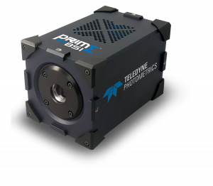
The Prime BSI back-illuminated sCMOS allows the Frischknecht lab to image up to 20x faster with the same signal to noise ratio. With the increased sensitivity, lower exposure times allow for an increase in speed which produces a more accurate image of multiple channels, enhancing the ability to track individual Plasmodium parasites more reliably. This also means that their movement can be studied more accurately. Jessica told us, "Weak fluorescent proteins, which had been impossible to observe with our previous camera, can now be readily visualized."
In addition to the increase in speed, the Prime BSI has a much larger field of view resulting in the detection of nearly 3x more cells. This substantiates the produced data, solidifies statistics and makes the quality of scientific output much higher. In addition, this reduces the time needed to perform experiments, which reduces time at the microscope.

Learn More About The Prime BSI