Imaging Live Cell Exocytosis at the Central Laser Facility
Mr. Benji Bateman, Dr. Lin Wang
Central Laser Facility, Science and Technology Facilities Council, Swindon, UK
Background
Mr Bateman is a Link Scientist working with Dr. Lin Wang in the Central Laser Facility (CLF), which contains everything from lasers the size of a room to compact benchtop lasers. Mr Bateman uses lasers with microscopes to do fluorescence imaging for the academic community, who bid for time at the CLF for their research. Mr Bateman and others at the CLF need to constantly keep up to date with the latest imaging technologies in order to best enable a wide range of imaging needs.
The CLF caters to many different applications, including super-resolution techniques such as Stochastic Optical Reconstruction Microscopy (STORM) for extremely high-resolution imaging, in very low imaging photon budgets, which need detectors capable of photon counting for STORM reconstruction, especially under cryogenic conditions.
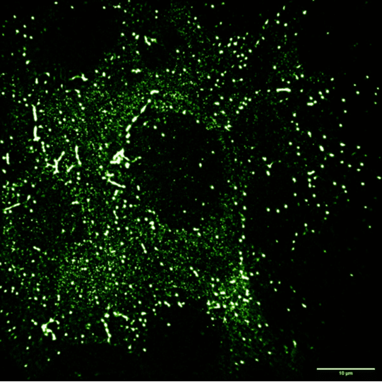
Figure 1: Chinese Hamster Ovary (CHO) cells transfected with wild-type EGFR labelled with 5 nM
EGF- Alexafluor488. TetraSpeck fiducial markers added and imaged at cryogenic temperatures.
Challenge
These CLF STORM imaging systems previously used EMCCD technologies, but these ran into issues with the dynamic range as Dr. Bateman outlined, "We're looking at samples in liquid nitrogen vapour and they bounce and wobble around, in order to correct for that we use bright TetraSpeck beads as fiducial markers, these are a lot brighter than the samples we measure and the well-depth of EMCCDs weren't sufficient to capture all of the fluorescent signals, we needed something with a larger dynamic range for the beads and the STORM information."
Mr. Bateman also mentioned they would benefit from a larger pixel for greater sensitivity, "We did try using a traditional CMOS with 6.5 µm pixels and but the sampling rate was mismatched to our choice of the microscope objective and solid immersion lens combinations, leading to overall magnifications up to 450x. We require the best STORM measurement precision possible in order to answer the biological questions our user groups have under investigation. This is only achievable with correctly sampled fluorescence point spread functions and high detector quantum efficiency. The Prime 95B met both of these requirements and currently supports all of our cryogenic user groups with their experiments."
We like the 95% quantum efficiency, the 11 µm pixel size, and the speed advantage, there isn’t really a good reason for us to use EMCCD any more with our cryogenic applications.
Mr. Benji Bateman, Dr. Lin Wang
Solution
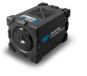
The Prime 95B CMOS meets the needs of super-resolution imaging systems while improving on previous EMCCD solutions in dynamic range, speed, field of view, and resolution while maintaining high sensitivity thanks to maximizing signal collection and minimizing noise.
Mr. Bateman spoke about the benefits of the Prime 95B, "We like the 95% quantum efficiency, the 11 µm pixel size, and the speed advantage are all benefits for us, there isn't really a good reason for us to use EMCCD any more... Another bonus of [the Prime 95B] would be the enhanced field of view and the larger number of pixels, especially compared to an EMCCD which has a very small chip size... We've coupled two 95Bs into a Cairn TwinCam using the full chip size for each channel.
Learn More About The Prime 95B
