Optical Fiber Photometry
Dr. Priya Rajasethupathy
Laboratory of Neural Dynamics and Cognition,
The Rockefeller University
Background
Dr. Priya Rajesethupathy and colleagues in the Rockefeller University Laboratory of Neural Dynamics and Cognition are interested in the process of memory. Integrating approaches ranging from genomics, transcriptomics, optogenetics and imaging, they aspire to address memory formation and recall at scales ranging from the synapse through to collections of hundreds to thousands of neurons and finally animal behavior.
The hippocampus region of the brain has long been thought as the place where memories form, but Dr. Rajesethupathy recently showed that a bundle of neurons that connect the hippocampus to the pre-frontal cortex is important for retrieving memories

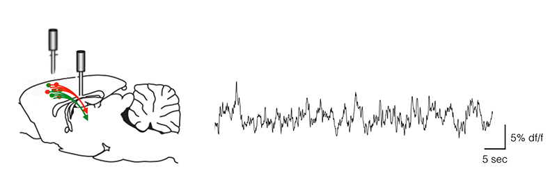
RightPlot of the change in fluorescence over the initial fluorescence acquired with thePrime 95Bfrom the nerve terminals, which previously had been too weak to detect reliably by photometry, is illustrated. Using the Prime 95B, dF/F values reaching 10% or greater can reliably be detected.
Challenge
Dr. Rajesethupathy's lab often measure neuronal activity of deep brain regions in a live mouse behaving in a virtual 3-dimensional world. To obtain neural activity information from select cell bodies and nerve bundle termini, fiber optics are implanted to deliver excitation light and recover fluorescence changes from neural activity sensors in brain regions of interest.
Collecting signals from cell-bodies can be easier than collecting signals from nerve bundle terminals. The sensor they use is cytoplasmic and because nerve terminal bundles contain less volume, they contain less sensor. This means they tend to have signals that are too weak to detect by traditional PMTs and sCMOS sensors. They also aim to record signals at speeds relevant to neuronal activation, which can be as fast as milliseconds.
The low noise and high quantum efficiency of the Prime 95B allows us to get good signal to noise and fast sampling from even sparsely innervated nerve termini
Dr. Priya Rajasethupathy
Solution
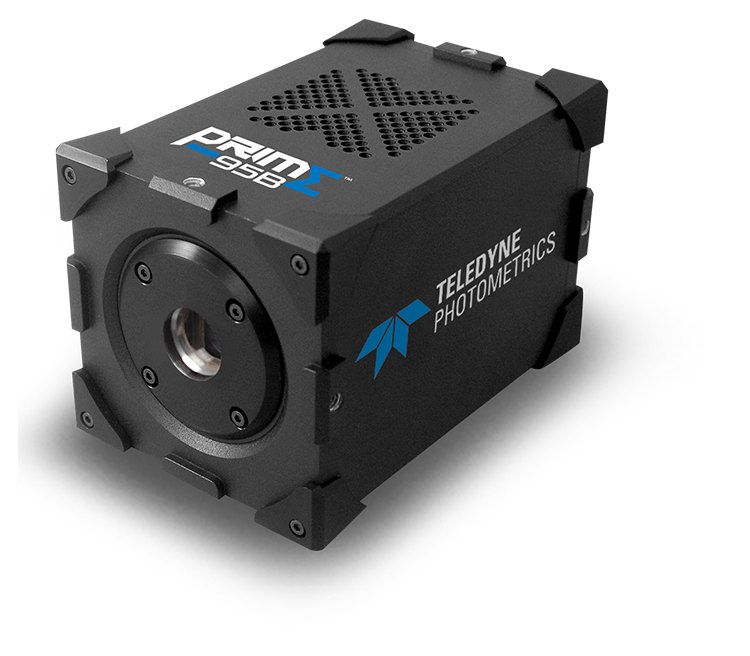
The Neural Dynamics and Cognition lab now uses the Teledyne PhotometricsPrime 95B sCMOS camerato collect the light through fiber optics from various regions in the brain.
Fibers collect light from characterized regions but give up spatial information in the process. Previous camera solutions allowed good signal to noise and high temporal precision from fibers implanted near groups of cell bodies, but not from regions rich in axonal synapses.
Dr. Rajasethupathy told us "Using the Prime 95B we collect these weak signals from nerve terminals very efficiently with good signal to noise (as shown in Figure 1). This reveals new biology that we are excited to explore."

Learn More About The Prime 95B