Sub-cellular Oblique Plane Microscopy
Dr. James Manton
MRC Laboratory of Molecular Biology, University of Cambridge, UK
Background
Dr. James Manton develops new microscopy techniques in the MRC Laboratory of Molecular Biology, Cambridge. A recent development project involves an oblique plane microscope (OPM) with a Mr. Snouty solid-immersion objective, which combines the speed and efficient illumination of light-sheet with the ease of use of a traditional inverted microscope. Dr. Manton aims to do multicolor imaging at higher speeds than existing light-sheet systems allow while maintaining the standard sample presentation of an inverted microscope.
This light sheet imaging system has been designed for a wide variety of samples, including highly photosensitiveDictyosteliumslime molds, T-cells, mouse fibroblasts, and other samples that require the gentle illumination of light-sheet.
Dr. Manton told us about their new light-sheet imaging system, "Because we are using a galvo mirror rather than moving the stage through the 3D acquisition, we can go five, ten times faster. This is particularly nice because a lot of the processes we want to look at, e.g.,Dictyosteliumare extremely fast. On our traditional light-sheet microscope we can acquire one volume a second, but here we are aiming for up to ten."

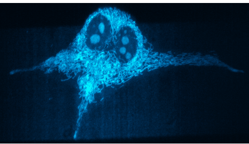
image shows a cyan stain formitochondria within a single cell.
Challenge
As the OPM will involve imaging volumes at high speed, a suitably high-speed camera is needed to capture all the light coming from the microscope in real-time. This kind of high-speed imaging requires low exposure times, combined with the low illumination level of light-sheet and the highly photosensitive samples, a highly sensitive camera is also needed to maximize the use of the photon budget.
In addition, this imaging system has a fixed magnification (55.7x), meaning a specific pixel size is required in order for optimal Nyquist sampling and high-resolution imaging.
Dr. Manton also mentioned some issues with previous sCMOS cameras, "we had issues with gain variation on previous sCMOS solutions - when we used an ROI to look at a single cell the non-uniformity in the gain became really clear at these low signal levels. This also made deconvolution trickier."
A new sCMOS imaging solution would need to have both low noise levels and no patterns or artifacts on the sensor, in order to have high sensitivity and reliable post-processing.
The [Prime BSI Express] gives us better gain variation and raw image quality compared to other sCMOS-style cameras.
Dr. James Manton
Solution
The Prime BSI Express is a flexible, reliable and powerful sCMOS camera, featuring high speed combined with high sensitivity.
Dr. Manton described his experience with the Prime BSI Express "We knew we wanted an sCMOS-style camera because of their speed, pixel size, and sensitivity... The gain variation appears to be a lot flatter on the Prime BSI Express compared to typical sCMOS, resulting in superior raw image quality."
"We have two [Prime BSI Express] on separate light paths split by a dichroic mirror. The system is run with MicroManager and the Photometrics device adaptor works just as expected, with nice, flat images at low light levels."
The Prime BSI Express has a clean, pattern-free bias and low noise CMS mode. Combining this with the near-perfect 95% quantum efficiency results in a highly sensitive camera that can run at 95 fps across the full sensor, allowing for high-speed imaging across large volumes.

Learn More About The Prime BSI Express
Download This Customer Story