Light Sheet Microscopy at University of St. Andrews
Prof. Kishan Dholakia, Dr. Stella Corsetti
Optical Manipulation Group, School of Physics and Astronomy, University of St. Andrews
Background
The Optical Manipulation Group at the University St. Andrews, led by Prof. Kishan Dholakia, looks at the science of light in terms of physics and biomedical research, fundamental and applied, in order to further the agenda for healthcare applications.
Prof. Dholakia wishes to take very high-quality images, in his terms: “A large field of view with high resolution, very quickly, very accurately, using the lowest possible photon dose.” For these reasons, the group uses light-sheet microscopy for the ability to take images with a lower light dose over a large field of view, with an aim to shed light on numerous biological processes.

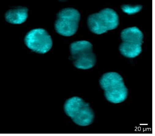
Figure 1: Taken with the Iris 15, this image shows mouse embryos at the 2-cell stage, stained with mitochondrial stain JC1, and imaged using an open-top light-sheet setup with a resolution of 900 nm.
Challenge
With live samples, the need to avoid photodamage is necessary in order to avoid affecting biological functions, when samples need to be imaged for days. Prof. Dholakia outlined some other issues: “We had challenges in how do we create the field of view, and how do we do Nyquist sampling across this field of view succinctly and accurately.”
Previous CMOS solutions were not able to deliver the desired image quality, meaning that a new solution was needed in order to achieve the large field of view images while retaining enough sensitivity to use lower light levels and preserve live samples for longer, as well as the ability to image with high resolution at low magnifications.
What we found exciting was the very small pixel size [of the Iris 15], which means we can retain the resolution but still get the big picture.
Prof. Kishan Dholakia, Dr. Stella Corsetti
Solution
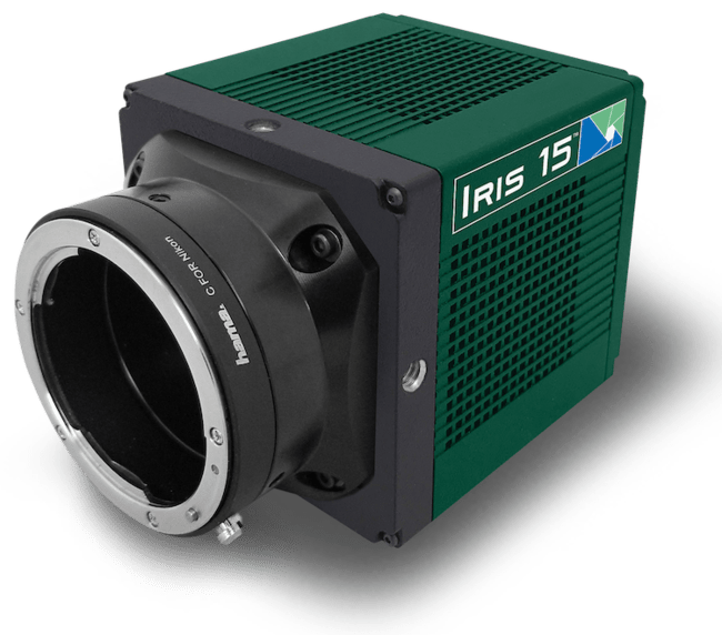
Prof. Dholakia and the team are now using the Iris 15 sCMOS, with features a rectangular 25 mm field of view, ideal for light sheet imaging. As Prof. Dholakia stated: “What we found exciting was the very small pixel size, which means we can retain the resolution but still get the big picture.” The small 4.25 μm pixel size allows for high resolution at low magnifications with Nyquist sampling, in combination with the large field of view this makes the Iris 15 an ideal solution over other sCMOS cameras.
Postdoc Dr. Stella Corsetti also outlined some of the advantages of the Iris 15: “The Iris15 camera gives us the advantage to Nyquist sample the resolution over a wide field of view due to the small pixel size.”
Prof. Dholakia also liked the camera dimensions: “The size of the camera is quite advantageous, we have been using other sCMOS cameras but the smaller physical size of the Iris 15 is important, we are interested in building platforms of microscopes that can fit on small breadboards, space is a premium and the Iris 15 fills a need there.”
