Live Vesicle Trafficking
Dr George Sirinakis, Senior Research Associate
St Johnston Lab, part of the Wellcome Trust/Cancer Research UK Gurdon Institute, University of Cambridge, UK
Background
The St Johnston Lab is primarily interested in how cells become asymmetric so that they can perform distinct functions on opposite sides of the cell. This is known as cell polarity.
In particular, the lab are interested in epithelial cells, which form the sheets that line our organs. Epithelial cells must have an apical side which contacts the lumen, for example the inside of your small intestine, lateral membranes where cell-cell contacts are made, and a basal surface that maintains contact with the extracellular matrix.
Loss of polarity, especially of epithelial cells, is a hallmark of cancer. In this project the lab aims to image the movement of vesicles with cargo proteins destined for these different membranes.
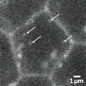
Figure 1 Drosophila epithelial cells. The arrows
indicate vesicles containing a cargo protein,
cell adhesion molecule Fasciclin III (FAS3) tagged
with Halo and labeled with SiR dye.
Challenge
The St Johnston Lab are aiming to image fast moving, small vesicles that contain a limited number of cargo proteins which results in a very low fluorescence signal. What is more, the lab are imaging in tissue, which causes further reductions in brightness due to aberrations. Finally, the tissue needs to be kept alive for imaging so the laser power must be kept to a minimum to reduce any phototoxic effects.
The high sensitivity and low read noise of the Prime BSI is transformative in these experiments as it enables us to use low levels of excitation power and significantly reduce photo-damage to the tissue.
Dr George Sirinakis
Solution
The Prime BSI back-illuminated sCMOS camera was a great solution for the St. Johnston Lab, combining high, 95% quantum efficiency and low read noise with the fast speed expected of an sCMOS device.
Dr Sirinakis shared with us, "The high quantum efficiency and dynamic range of the Prime BSI enables us to visualize even the dimmest vesicles that contain limited numbers of cargo molecules and track them with high speeds in live tissue."
Dr Sirinakis went on to say, "The high sensitivity and low read noise of the Prime BSI is transformative in these experiments as it enables us to use low levels of excitation power and significantly reduce photo-damage to the tissue. With the Prime BSI we are now able to ask more meaningful questions about vesicle trafficking and better understand cell polarity."
Learn More About the Prime BSI
Download This Customer Story