Live Cell Fluorescence Microscopy
Christof Osman, PhD, Professor
Ludwig-Maximilian University, München, Germany
Department of Biology II, Faculty of Biology, Cell and Developmental Biology
Background
The lab of Prof. Osman's at the Ludwig-Maximilian University in München, Germany is interested in understanding the power plants of cells - mitochondria - and the way their functionality and network activity is maintained during cell division. The underlying process of ensuring that the mitochondrial DNA is correctly distributed between cells undergoing cell division so that cells can produce the required proteins, is the lab's goal. The team developed a tool which allows them to track in yeast mitochondrial DNA, in the least-possible invasive manner, with high-speed live-cell fluorescence microscopy.
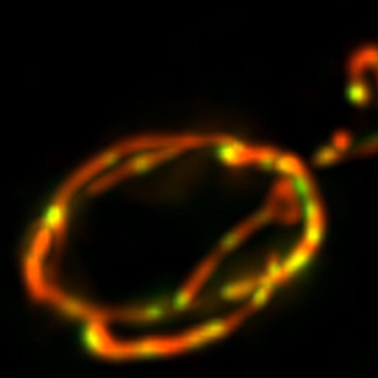
Maximum intensity projection of 45 planes showing live yeast mitochondria
labelled with mKate2 and mitochondrial DNA with tandem-mNeonGreen.
Image was taken with a Photometrics Prime 95B 25mm CMOS camera on a
Nikon Ti2 microscope.
Challenge
The lab's research organism is yeast, which is notoriously challenging to image as samples are small and low in fluorescence. Moreover, as the process of cell division and the function of mitochondria is very sensitive to exposure with light (in particular with wavelength in the UV and blue end of the spectrum), the research team would like to use a light dose, as low as possible, for excitation. This allows them the ability to image samples under physiological conditions. Additionally, the events to be observed can be relatively rare, and therefore it is important to image as many cells as possible in one attempt to achieve optimal statistics.
The Prime 95B [Scientific CMOS] camera provides the required sensitivity that is needed to image our yeast cells with as little excitation light as possible.
Christof Osman
Solution
Prof. Osman is using two Prime 95B 25mm Scientific CMOS cameras with a new TwinCam which is suited for the 25mm diameter light path of the Nikon Ti2 microscope. "The Prime 95B camera provides the required sensitivity that is needed to image our yeast cells with as little excitation light as possible. This prevents phototoxic effects and allows us to maintain cells under physiological conditions throughout long imaging experiments," Prof. Osman shares. The TwinCam allows the ability to image two channels simultaneously without having the need to switch filters in-between which leads to losing temporal resolution.
Learn More About the Prime 95B