Whole Tissue Calcium Imaging
Dr. Marcel Hörning
Institute of Biomaterials and Biomolecular Systems, University of Stuttgart, Germany
Background
Dr. Marcel Hörning is a physicist and bioengineer, the Principal Investigator of the Biobased Materials Group, led by Prof. Ingrid Weiss at the University of Stuttgart. Dr. Hörning recently obtained funding from the DFG for research into electro-mechanical wave formations in cardiac tissue.
Dr. Hörning described his recent work, "We have a model cardiac system involving re-engineered ex vivo primary tissue cultures of the heart, we grow these cardiac tissues in the lab and observe the electro-mechanical waves across the tissue. We have action potentials, calcium signaling, and mechanical contraction, all synchronized and in patterns such as spiral waves and alternans."
"We found a simple method using Fourier transformation to visualize these patterns in real-time, with this Fourier transformation imaging (FFI) we can identify complex patterns of alternans using calcium imaging, membrane potential, and contraction patterns."

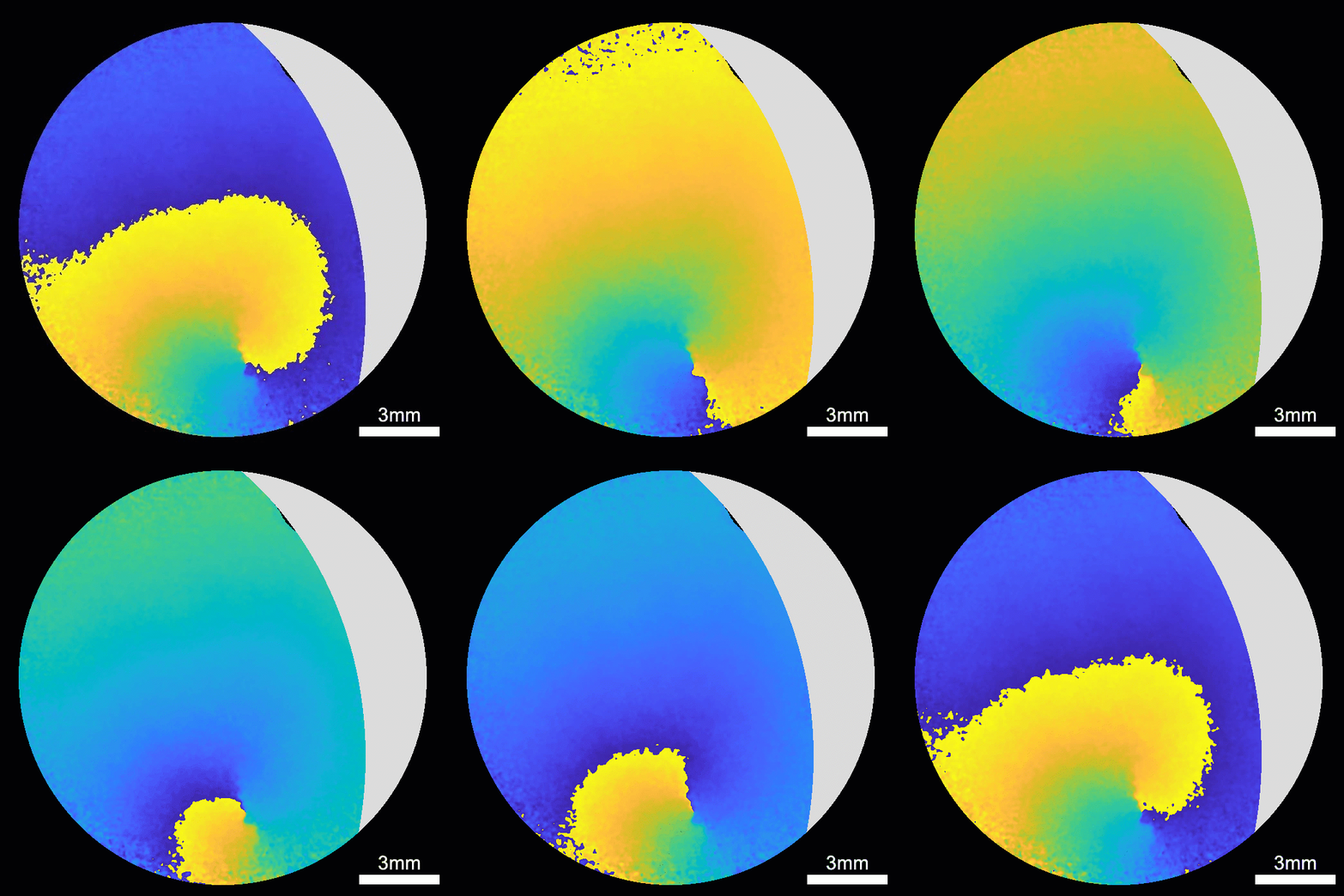
Figure 1: Spiral calcium waves across a piece of in vitro cardiac tissue, taken with the Kinetix. The image
shows a 1.5 cm diameter piece of tissue, imaged with a 2x lens and background(minimum intensity) subtraction.
The grey section contains no cells.Recorded by Julia Erhardt.
Challenge
Imaging functional activity over a large piece of tissue requires a camera with a high spatial and temporal resolution, high speed for functional activity recordings (calcium and membrane potential), and high spatial resolution for imaging morphology at a cellular level within the tissues.
Dr. Hörning explained his imaging challenges, "We want to image over a large field of view at a 2x magnification with sub-cellular resolution. We also need to capture at a high speed in order to resolve the signals. Essentially, we need a combination of high spatial and temporal resolution in order to detect the alternans patterns with FFI."
By obtaining high-quality structural and functional information from the cardiac tissue models, Dr. Hörning works to improve the model and increase the biological relevance to the in vivo situation.
Overall the Kinetix is a very impressive camera, the large field of view and high speed meet my need for high spatial and temporal resolution imaging.
Dr. Marcel Hörning
Solution
The Kinetix sCMOS is a powerful solution for both large format and high-speed imaging. The large field of view (29 mm diagonal) and small pixel (6.5 μm) allow for high-resolution imaging across a large sample, while the high acquisition speeds (500 fps across the full-frame) allow for easy capture of fast, dynamic events such as calcium waves and other functional cellular activity.
Dr. Hörning told us about his experience with the Kinetix, "We got support from Teledyne Photometrics the whole time, it all worked well and I didn't have any trouble. I'm happy it works so smoothly and I was able to get results."
"We use Sensitivity mode but would also be interested in optimizing the Speed mode as this 8-bit mode decreases the data file size. Overall, the Kinetix is a very impressive camera and meets the needs of my research."

Learn More About The Kinetix
Download This Customer Story