Organoid Light Sheet Imaging
Dr. Franziska Decker
Control Theory and Systems Biology Lab, ETH Zürich, Switzerland
Background
Dr. Franziska Decker aims to follow the differentiation of tissues and the development of organoids in 3D using a custom-built light sheet microscope. This work is mainly done with kidney organoids and gastruloids.
Experiments involve the light sheet imaging system in order to scan through layers of cells in an organoid. By taking z-stacks through the organoid at regular intervals over the long term, growth can be monitored. Due to the large size of the samples, more penetrative wavelengths are preferred, leading Dr. Decker to use near-infrared fluorescent proteins such as miRFP.

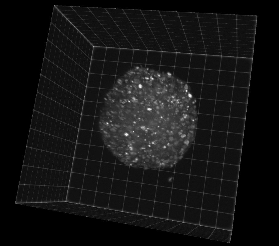
Figure 1: Image of a gastruloid taken with the Iris 15 on a light sheet microscopy system. The organoid is labeled
with a DNA marker miRFP-H2B, which emits in the far red.
Challenge
Dr. Decker outlined some of the challenges she faces when imaging, "Ideally we want to use light sheet microscopy to watch organoids grow up to a millimeter or more in size, for that we need a large field of view with enough resolution to be able to see the nuclei of the cells for tracking."
"We sometimes have 300-400 z-stack planes to image in order to get through the entire organoid, if we do that with three different channels it's also nice to have speed so imaging doesn't take a full hour."
A suitable solution for this light sheet application would require a large field of view, a small pixel size for high resolution and low magnification (16x), as well as speed in order to increase the throughput and perform more efficient multidimensional imaging of large samples over the long term.
So far every sample I have imaged fits into one field of view, and [the Iris 15] works great, it was easy to setup and image with.
Dr. Franziska Decker
Solution
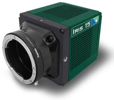
The Iris 15 is designed for large field of view, low magnification light sheet imaging, making it the ideal solution for this application. With a small 4.25 um pixel and a massive 25 mm field of view thanks to the 5056 x 2960 pixel array, the Iris 15 is a high-throughput sCMOS with very high resolution, all operating at 30 fps.
Dr. Decker told us about her experience with the Iris 15, "we talked to various companies and in the end, I decided on the Iris [15], mainly because of the small pixel size and huge field of view, it is ideal for the imaging we want to do... so far I really like it and I'm happy with the images and the integration with Micro-Manager."
