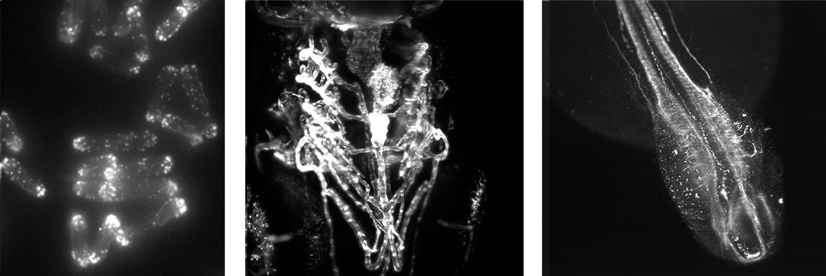
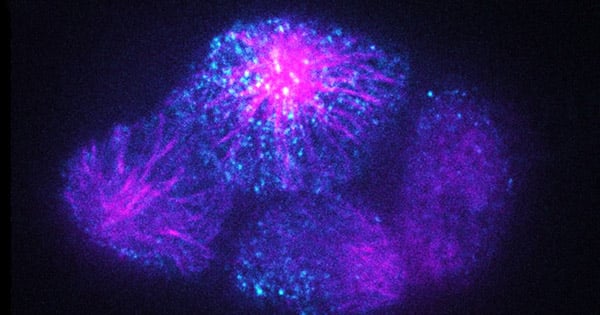
Light sheet microscopy enables scientists to overcome two major problems in modern microscopy. Namely, to image biological samples for much longer under physiologically relevant conditions than with conventional microscopy techniques and to image samples of considerable size in a more reasonable and relevant time frame.
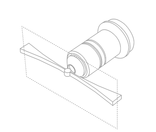
This is achieved by illuminating the sample with a sheet of light positioned perpendicular to the imaging axis and thereby illuminating only the plane in focus. As a result, out-of-focus structures will see less light and the sample is therefore less likely to develop phototoxic products.
Light sheet microscopy combines the best features of two worlds. Signal to noise will be enhanced when comparing it with widefield illumination. Acquisition speed will benefit from using a camera rather than the point scanner of a confocal.
Resources from the Learning Center
View All
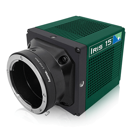
Iris 15
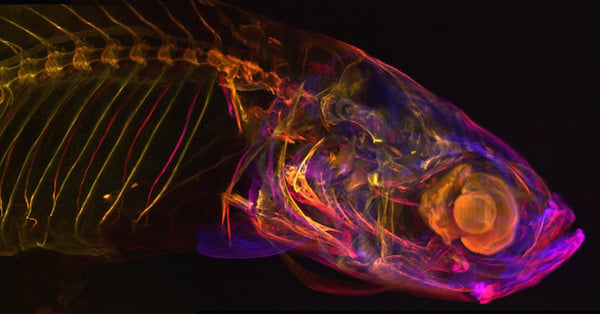
The Iris 15 is specifically designed for light sheet microscopy with a massive 25 mm diagonal field of view and small 4.25×4.25 µm pixels. For reasons of geometry, light sheet microscopy is typically performed with low magnification objectives which requires a smaller pixel for Nyquist sampling. The small pixels of the Iris 15 give greatly improved resolution over typical sCMOS devices to maximize the level of detail detected in the sample. The range of light sheet microscopy techniques and geometries is staggering and for many, having a large field of view is of great benefit. A larger field of view camera allows larger samples to be imaged without needing to stitch multiple images together or, for smaller samples, the large field of view can be split to perform multichannel imaging easily on the same sensor. The Iris 15 also features Programmable Scan Mode, which allows the user control over the rolling shutter exposure and read-out functionality of the sensor to allow optimization around applications that require control over the line time.
View Product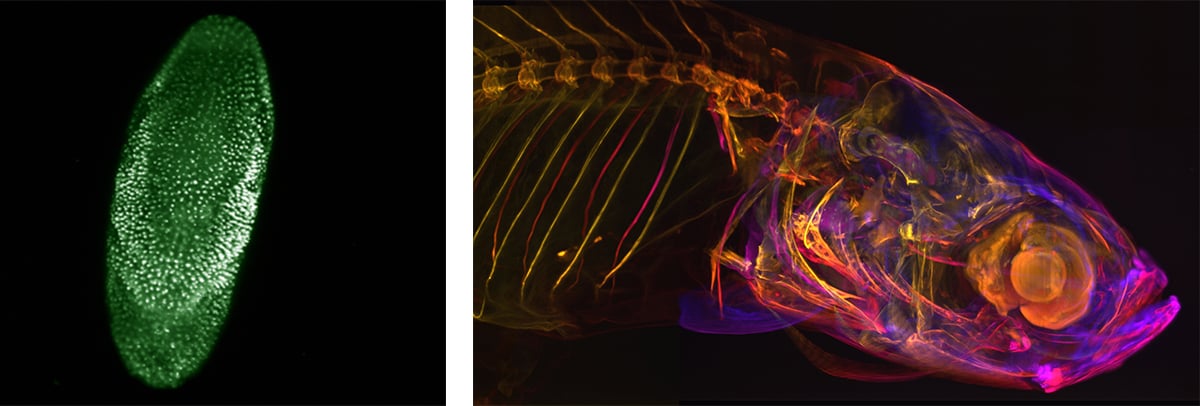
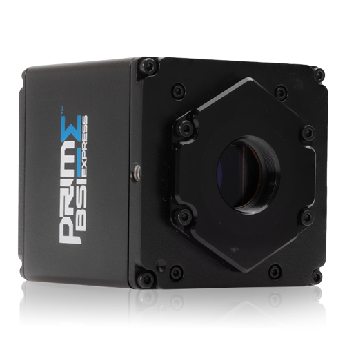
Prime BSI Express
High sensitivity, 95% quantum efficient, sCMOS camera with 6.5 µm pixels, 1.0 e– read noise and a full-frame framerate of 95 fps. The high quantum efficiency and low read noise combined with the balanced 6.5 µm pixel size offers higher sensitivity than conventional sCMOS devices whilst maintaining the resolution, large field of view and high speed. When the Iris 15 isn’t enough, the Prime BSI Express provides the necessary boost in sensitivity and speed.
View Product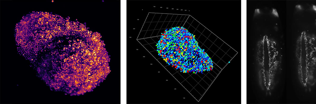
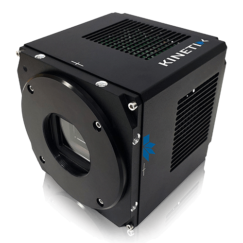
Kinetix
High sensitivity, 95% quantum efficient sCMOS camera with an incredibly high 500 fps full-frame speed and a massive 29.4 mm diagonal field of view. When speed is needed, the Kinetix significantly outperforms typical sCMOS devices. With a full-frame speed of 500 fps, light sheet microscopy techniques that rely on fast framerates such as SCAPE or lattice light sheet microscopy will be able to deliver more than ever before. For larger samples, the 29.4 mm square sensor of the Kinetix is designed to increase throughput, maximize the amount of data captured in a single frame and significantly speed up data acquisition.
View Product